Heat-Treatments Affect Protease Activities and Peptide Profiles of Ruminants’ Milk. 

Heat-Treatments Affect Protease Activities and Peptide Profiles of Ruminants’ Milk. 

Juliana A. S. Leite1Carlos A. Montoya1,2Simon M. Loveday1,2Evelyne Maes3Jane A. Mullaney1,2,4Warren C. McNabb1,4* and Nicole C. Roy1,4,5,6

Frontiers in Nutrition 2021

Proteases present in milk are heat-sensitive, and their activities increase or decrease depending on the intensity of the thermal treatment applied. The thermal effects on the protease activity are well-known for bovine milk but poorly understood for ovine and caprine milk. This study aimed to determine the non-specific and specific protease activities in casein and whey fractions isolated from raw bovine, ovine, and caprine milk collected in early lactation, and to determine the effects of low-temperature, long-time (63°C for 30 min) and high-temperature, short-time (85°C for 5 min) treatments on protease activities within each milk fraction. The non-specific protease activities in raw and heat-treated milk samples were determined using the substrate azocasein. Plasmin (the main protease in milk) and plasminogen-derived activities were determined using the chromogenic substrate S-2251 (D-Val-Leu-Lys-pNA dihydrochloride). Peptides were characterized using high-resolution liquid chromatography coupled with tandem mass spectrometry. The activity of all native proteases, shown as non-specific proteases, was similar between raw bovine and caprine milk samples, but lower (P < 0.05) than raw ovine milk in the whey fraction. There was no difference (P > 0.05) between the non-specific protease activity of the casein fraction of raw bovine and caprine milk samples; both had higher activity than ovine milk. After 63°C/30 min, the non-specific protease activity decreased (44%; P > 0.05) for the bovine casein fraction only. In contrast, the protease activity of the milk heated at 85°C/5 min changed depending on the species and fraction. For instance, the activity decreased by 49% for ovine whey fraction, but it increased by 68% for ovine casein fraction. Plasmin and plasminogen were in general inactivated (P > 0.05) when all milk fractions were heated at 85°C/5 min. Most of the peptides present in heat-treated milk were derived from β-casein and αS1-casein, and they matched the hydrolysis profile of cathepsin D and plasmin. Identified peptides in ruminant milk samples had purported immunomodulatory and inhibitory functions. These findings indicate that the non-specific protease activity in whey and casein fractions differed between ruminant milk species, and specific thermal treatments could be used to retain better protease activity for all ruminant milk species.

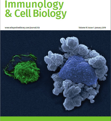
Early‐life exposure to gut microbiota from disease‐protected mice does not impact disease outcome in type 1 diabetes susceptible NOD mice

Early‐life exposure to gut microbiota from disease‐protected mice does not impact disease outcome in type 1 diabetes susceptible NOD mice

Immunology and cell biology  

Abstract

The microbial community making up the gut microbiota can profoundly influence intestinal homeostasis and immune system development, and is believed to influence the development of complex diseases including type 1 diabetes (T1D). T1D susceptible nonobese diabetic (NOD) mice have been shown to harbor a distinct microbiota to disease-protected mice. We hypothesized that the T1D susceptible genetic background of NOD mice would be resistant to the introduction of a C57BL/6-derived microbiota. NOD and C57BL/6 mice were cohoused either continually from birth, from birth until weaning or from weaning onwards, allowing transfer of microbiota between the mice. Cohousing NOD with C57BL/6 mice from before birth, resulted in moderate changes to the gut microbiota, whereas initiating cohousing at weaning only led to minimal changes. Terminating cohousing at weaning reduced the changes in the microbiota composition. However, diabetes onset was not significantly delayed and there was no reduction in intestinal inflammation or the proportion of regulatory T cells in the cohoused NOD mice. However, insulin but not islet-specific glucose-6-phosphatase catalytic subunit-related protein-specific CD8+ T cells were reduced by cohousing suggesting an epitope-specific modulation of the autoreactive response by the gut microbiota. These results suggest that the T1D susceptible genetic background of the NOD mouse was resistant to the introduction of a C57BL/6-derived microbiota.

Intestinal Metaproteomics Reveals Host-Microbiota Interactions in Subjects at Risk for Type 1 Diabetes

Intestinal Metaproteomics Reveals Host-Microbiota Interactions in Subjects at Risk for Type 1 Diabetes

Abstract

OBJECTIVE Dysbiosis of the gut microbiota has been linked to disease pathogenesis in type 1 diabetes, yet the functional consequences to the host of this dysbiosis are unknown. We investigated the functional interactions between the microbiota and the host associated with type 1 diabetes disease risk.

RESEARCH DESIGN AND METHODS We performed a cross-sectional analysis of stool samples from subjects with recent-onset type 1 diabetes (n = 33), islet autoantibody–positive subjects (n = 17), low-risk autoantibody-negative subjects (n = 29), and healthy subjects (n = 22). Metaproteomic analysis was used to identify gut- and pancreas-derived host and microbial proteins, and these data were integrated with sequencing-based microbiota profiling.

RESULTS Both human (host-derived) proteins and microbial-derived proteins could be used to differentiate new-onset and islet autoantibody–positive subjects from low-risk subjects. Significant alterations were identified in the prevalence of host proteins associated with exocrine pancreas output, inflammation, and mucosal function. Integrative analysis showed that microbial taxa associated with host proteins involved in maintaining function of the mucous barrier, microvilli adhesion, and exocrine pancreas were depleted in patients with new-onset type 1 diabetes.

CONCLUSIONS These data support that patients with type 1 diabetes have increased intestinal inflammation and decreased barrier function. They also confirmed that pancreatic exocrine dysfunction occurs in new-onset type 1 diabetes and show for the first time that this dysfunction is present in high-risk individuals before disease onset. The data identify a unique type 1 diabetes–associated signature in stool that may be useful as a means to monitor disease progression or response to therapies aimed at restoring a healthy microbiota.


Type 1 diabetes susceptibility alleles are associated with distinct alterations in the gut microbiota

Type 1 diabetes susceptibility alleles are associated with distinct alterations in the gut microbiota

Abstract Background: Dysbiosis of the gut microbiota has been implicated in the pathogenesis of many autoimmune conditions including type 1 diabetes (T1D). It is unknown whether changes in the gut microbiota observed in T1D are due to environmental drivers, genetic risk factors, or both. Here, we have performed an analysis of associations between the gut microbiota and T1D genetic risk using the non-obese diabetic (NOD) mouse model of T1D and the TwinsUK cohort. Results: Through the analysis of five separate colonies of T1D susceptible NOD mice, we identified similarities in NOD microbiome that were independent of animal facility. Introduction of disease protective alleles at the Idd3 and Idd5 loci (IL2, Ctla4, Slc11a1, and Acadl) resulted in significant alterations in the NOD microbiome. Disease-protected strains exhibited a restoration of immune regulatory pathways within the gut which could also be reestablished using IL-2 therapy. Increased T1D disease risk from IL-2 pathway loci in the TwinsUK cohort of human subjects resulted in some similar microbiota changes to those observed in the NOD mouse. Conclusions: These findings demonstrate for the first time that type 1 diabetes-associated genetic variants that restore immune tolerance to islet antigens also result in functional changes in the gut immune system and resultant changes in the microbiota. Keywords: Gut microbiota, Type 1 diabetes, Genetic susceptibility, Interleukin-2 pathway, Autoimmunity


Modulation of the microbial fermentation in the gut by fermentable carbohydrates

Modulation of the microbial fermentation in the gut by fermentable carbohydrates

Abstract

This review considers fermentable carbohydrates and their role in maintaining health through their availability as fuel for the gut microbiota. The microbiota possesses remarkably diverse function, and is likely modifiable by diet. Therefore a diet rich in varied fermentable carbohydrates such as dietary fibre, glycosylated polyphenolics, glucosinolates and other plant glycans, applied in a sustained fashion may promote microbial diversity leading to improved health. This may be achieved by increasing the flexibility of the microbiota’s capability to interact with diverse dietary environments, or via increasing production of short chain fatty acids (SCFAs) from the fermentation of carbohydrates. A higher functional modular complexity is indicative of gut health, whilst SCFAs may reduce the risk of developing gastrointestinal disorders, cancer, and cardiovascular disease.

Lactic Acid Bacteria Convert Glucosinolates to Nitriles Efficiently Yet Differently from Enterobacteriaceae

Lactic Acid Bacteria Convert Glucosinolates to Nitriles Efficiently Yet Differently from Enterobacteriaceae

Abstract

Glucosinolates from the genus Brassica can be converted into bioactive compounds known to induce phase II enzymes, which may decrease the risk of cancers. Conversion via hydrolysis is usually by the brassica enzyme myrosinase, which can be inactivated by cooking or storage. We examined the potential of three beneficial bacteria, Lactobacillus plantarum KW30, Lactococcus lactis subsp. lactis KF147, and Escherichia coli Nissle 1917, and known myrosinase-producer Enterobacter cloacae to catalyze the conversion of glucosinolates in broccoli extract. Enterobacteriaceae consumed on average 65% glucoiberin and 78% glucoraphanin, transforming them into glucoiberverin and glucoerucin, respectively, and small amounts of iberverin nitrile and erucin nitrile. The lactic acid bacteria did not accumulate reduced glucosinolates, consuming all at 30–33% and transforming these into iberverin nitrile, erucin nitrile, sulforaphane nitrile, and further unidentified metabolites. Adding beneficial bacteria to a glucosinolate-rich diet may increase glucosinolate transformation, thereby increasing host exposure to bioactives.

Abstract Image

Bacterial Polyhydroxyalkanoate Granules: Biogenesis, Structure, and Potential Use as Nano-/Micro-Beads in Biotechnological and Biomedical Applications

Bacterial Polyhydroxyalkanoate Granules: Biogenesis, Structure, and Potential Use as Nano-/Micro-Beads in Biotechnological and Biomedical Applications

Abstract

Polyhydroxyalkanoates (PHAs) are naturally occurring organic polyesters that are of interest for industrial and biomedical applications. These polymers are synthesized by most bacteria in times of unbalanced nutrient availability from a variety of substrates and they are deposited intracellularly as insoluble spherical inclusions or PHA granules. The granules consist of a polyester core, surrounded by a boundary layer with embedded or attached proteins that include the PHA synthase, phasins, depolymerizing enzymes, and regulatory proteins. Apart from ongoing industrial interest in the material PHA, more recently there has also been increasing interest in applications of the PHA granules as nano-/micro-beads after it was conceived that fusions to the granule associated proteins (GAPs) provide a way to immobilize target proteins at the granule surface. This review gives an overview of PHA granules in general, including biogenesis and GAPs, and focuses on their potential use as nano-/micro-beads in biotechnological and biomedical applications.